Mapping Aging Brain Networks with the Groundbreaking Atlas55+
Tuesday, January 5, 2021
Brain atlases of various kinds have existed for decades, each mapping various areas and centers of the brain. But up until now, most of these atlases created by various researchers have used subjects in early adulthood (typically 18 to 35 years old) for their studies.
Yet older individuals represent 15% of the United States population, and that segment is expected to continue growing significantly through 2050. This left a vacuum in the research arena when it came to looking at age-related brain changes and diseases, since no atlas for that age group existed, undermining the validity and reliability of neuroimaging research when it came to older adults.
That is what makes the Atlas55+ brain atlas, mapped by the
Boys Town Brain Architecture, Imaging and Cognition Lab and its affiliates, so important. Now scientists researching brain and cognition changes in later adulthood have a reliable atlas of the brain networks that has been created using functional MRIs of healthy individuals between the ages of 55 and 95.
“By providing the first age-adapted brain atlas for late adulthood to the scientific community, this work has the potential to reveal how dysfunction of the brain networks contributes to neurodegenerative conditions like dementia," reported
Gaelle Doucet, Ph.D., Director of the Brain Architecture, Imaging and Cognition Lab at Boys Town National Research Hospital®.
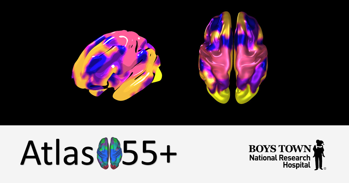
This brain atlas identified five major networks. The study conducted by Dr. Doucet found that three of these networks, the default-mode network (DMN), which is involved in internal-related functions such as thought generation and memory; the executive central network (ECN), which supports working memory, and the salience network (SAL), which helps the transition between different cognitive activities, showed the most changes in functional integrity in older adults, compared to younger adults.
These three networks “support high-order cognitive activity such as memory, attention and any type of mental activity that helps you work, live and think correctly," explained Dr. Doucet. “Our work showed that these three networks are particularly vulnerable to aging."
Though further research is needed, this new atlas of the aging brain may help explain why some adults struggle with cognitive decline as they age. This research suggests that interventions to prevent or attempt to reverse cognition loss should focus on the DMN, the ECN and the SAL since these are the major networks that lose functional integrity with aging.
Atlas55+ has been published and is available for other researchers to consult and use.
“We are hoping that other neuroscientists in the field of aging will use Atlas55+ as they conduct research with older populations," said Dr. Doucet. “Previously they only had atlases based on younger populations, which may create bias. Now there is an atlas available for working with studies of Alzheimer's, dementia and other age-related cognitive declines."
The end goal for Atlas55+ is that it can be used as a reference for any population above the age of 55 and that it will be able to aid in the diagnosis of neurodegenerative disorders such as mild cognitive impairment (MCI) or Alzheimer's disease by providing a comparative baseline for what a healthy aging brain networks look like.
The Atlas55+ research study was funded by the National Institute on Aging (NIA), which is part of the National Institute of Health, the research agency of the DHHS. NIA's mission is to sponsor biomedical, behavioral and social research nation-wide to improve the health of older adults.
“We know that age affects the brain throughout life, so my goal is to eventually take the same type of approach with children," said Dr. Doucet. “We need to create these same types of normative reference templates in children since none currently exist. I envision this eventually being done with 10- to 18-year-olds since younger children prove more difficult to get accurate brain scans from."
Researchers wishing to view the published article in the journal
Cerebral Cortex, published by Oxford Academic Journals, should use the following link:
academic.oup.com/cercor/article/31/3/1719/5981728.